The Effect of Polybutylcyanoacrylate Nanoparticles as a Protos Delivery Vehicle on Dental Bone Formation
圖文摘要說明
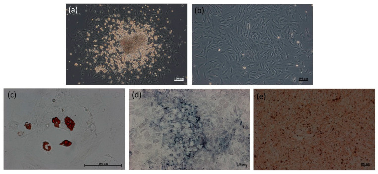
本組顯微鏡圖像便展示了評估此新型載鍶奈米材料促進骨生成能力的關鍵實驗結果:圖(a)與(b)呈現了從組織中分離出的幹細胞,顯示其具有典型的集落形成能力與紡錘狀形態;而後續的誘導分化實驗中,圖(d)呈現的深藍色「鹼性磷酸酶」(ALP)染色與圖(e)中棕紅色的「茜素紅」(Alizarin Red)鈣化結節染色,分別證實了骨生成早期與晚期的標誌性活動。與此同時,圖(c)中的「油紅O」染色(紅色脂肪滴)作為對照組,驗證了這些細胞具備多分化潛能的幹細胞特性。綜合而言,這一系列圖像有力地證明了此載鍶奈米載體能成功誘導幹細胞分化為功能性的骨細胞,為其應用於改善植牙骨整合提供了重要的體外實驗依據。
Abstract
Background: Dental implants are commonly used for missing teeth, for which success depends heavily on the quality of the alveolar bone. The creation of an ideal implant site is a key component in shortening the treatment time, which remains clinically challenging. Strontium ranelate (Protos) is an anti-osteoporotic agent which has previously been used to promote bone formation, however the systemic use of Protos has been linked to serious cardiovascular and venous thromboembolic events, thus local delivery strategies may be better suited for this purpose. In this study, a biodegradable, and biocompatible nanocarrier "polybutylcyanoacrylate" (PBCA) loaded with strontium was constructed and its ability to promote bone formation was assessed.
Methodology: PBCA nanoparticles loaded with strontium (PBCA-Sr NPs) were synthesized using the emulsion polymerization method, and their physical properties (zeta potential, size and shape) and entrapment efficiency were characterized. Committed MSCs (osteoblasts) were derived from the differentiation of cultured rat mesenchymal stem cells (MSC), which were tested with the PBCA-Sr NPs for cytotoxicity, inflammatory response, bone formation and mineralization. Scanning electron microscopy was performed following a 7-day treatment of PBCA-Sr NPs on decellularized procaine mandibular bone blocks grafted with osteoblasts.
Results: Spherical PBCA-Sr NPs of 166.7 ± 2.3 nm, zeta potential of -1.15 ± 0.28 mV with a strontium loading efficiency of 90.04 ± 3.27% were constructed. The presence of strontium was confirmed by energy-dispersive X-ray spectroscopy. Rat committed MSCs incubated in PBCA-Sr NPs for 24 hrs showed viabilities in excess of 90% for concentrations of up to 250 ug/mL, the cellular expression of osteocalcin and alkaline phosphatase were 1.4 and 1.3 times higher than the untreated control, and significantly higher than those treated with strontium alone. Bone formation was evident following osteoblast engraftment on the decellularized procaine mandibular bone block with PBCA-Sr NPs, which appeared superior to those treated with strontium alone.
Conclusion: Treatment of committed MSCs with PBCA-Sr NPs showed higher expression of markers of bone formation when compared with strontium alone and which corresponded to greater degree of bone formation observed on the 3-dimensinal decellularized procaine mandibular bone block. Further quantitative analysis on the extent of new bone formation is warranted.
Keywords: bone formation; nanotechnique; polybutylcyanoacrylate; strontium.
瀏覽數: